Bone Brain Connection
October 29, 2009 Written by JP [Font too small?]The connection between mental health and the condition of the physical body is often neglected in modern medicine. One example is the way conventional doctors generally treat bone loss, otherwise known as osteopenia (minor loss of bone density) and osteoporosis (significant loss of bone mineralization). The typical advice given is to take the “recommended daily allowance (RDA)” of supplemental calcium and Vitamin D, hormone replacement therapy and a select group of medications that attempt to “harden” the bones. But one suggestion that I’ve never heard from an allopathic physician is to practice stress management as a way of protecting the skeletal system.

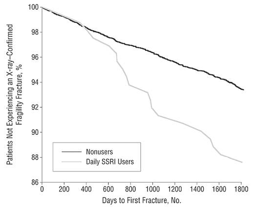
Emerging evidence suggests that excess production of cortisol, the body’s primary stress hormone, may contribute to poor bone status in older and younger populations alike. It also appears that this proposed connection is a risk factor for both genders. But there’s even more to the story. Simply treating stress with medications doesn’t appear to improve skeletal health. In fact, some of the drugs used to address anxiety and depression may actually make things worse.
A new study in the Journal of Clinical Endocrinology and Metabolism provides a powerful example of the correlation between “hypercortisolism” and poor bone status. This Italian examination looked for an association between mildly elevated stress hormones and low bone density. 287 patients with subclinical hypercortisolism (SH) were compared to 194 healthy volunteers. Both groups contained a broad cross section of men and women of varying ages. X-rays were taken of all the participants at two relevant locations (femoral neck and lumbar spine). The researchers determined that those with high cortisol had dramatically lower bone density in the femoral neck and lumber spine regions. There was also a more than 3 times greater risk of fractures in the patients with SH. This additional risk was not influenced by factors relating to age, gender or menopausal status. (1)
There have been several other trials from 2009 that support the osteroporotic/stress connection. Each of the studies focuses on a different population group – adult men, overweight adolescents and anorexic women. This is important to note because the negative effects of stress hormones on the skeletal system can apparently impact all of these disparate groups. Therefore, the possibility exists that this unwanted side-effect could also apply to the population at large.
- A 12 month observational trial was conducted on 88 men with subclinical hypercortisolism and 90 healthy subjects. Bone mineral density and cortisol levels were measured at the start and end of the study period. Femoral neck and lumbar spine bone status was dramatically lower in those with high cortisol levels. The lumber vertebrae were found to be particularly porous, which could explain the higher “prevalence of vertebral fractures” in this sensitive group. (2)
- Anorexia nervosa (AN) is an eating disorder that is characterized by excessive cortisol production. A recent experiment conducted at Massachusetts General Hospital and Harvard Medical School examined the role that “anxiety, depression and/or cortisol dysregulation contribute to low bone density”. As expected women with AN were found to produce larger quantities of cortisol and suffer from higher levels of anxiety and depression as compared to healthy volunteers. They also exhibited lower bone density at the hip and spine. The authors of the trial concluded that, “hypercortisolism is a potential mediator of bone loss and mood disturbance”. (3)
- The National Institutes of Health recently published a study that involved 137 overweight, adolescent boys and girls. Questionnaires, specialized interviews and various diagnostic tests were performed to determine bone density, eating patterns and psychological status in the youngsters. The researchers discovered a connection between the stress caused by “adolescent shape and weight concern” and lumbar spine bone density. The association between bone health and stress was determined via the measurement of urinary cortisol and x-rays. (4)
Anxiety and depression are typically associated with elevated cortisol levels. This may explain why these psychological conditions are now proposed as an independent and substantial risk factor for low bone density. A meta-analysis from September 2009 reviewed 23 studies that compared the bone status of depressed individuals to healthy volunteers. On average, depressed men and women demonstrated weaker bones in their forearms, hips and spine regions. Women appeared to be more vulnerable to the effects of psychological distress than men. An interesting side note is that premenopausal women were at greater danger for fractures than postmenopausal women. A separate study in the journal Bone also suggests a strong correlation between depression in premenopausal women and low bone density. The authors of that research recommend that such patients “should be investigated for osteroporosis”. (5,6,7,8)

In order to investigate how stress alters bone density, researchers have looked to a condition known as Cushing’s syndrome, which features “overt hypercortisolism” and can lead to “osteoporosis and fractures in up to 70% of cases”. In such circumstances, there is a dysfunction in the mechanisms by which bones are broken down and rebuilt. There may also be some indirect ways that cortisol exacts its damage, such as altering sex hormone levels and promoting calcium malabsorption. Certain medications can also inadvertently contribute to the problem. For instance, depressed patients given anxiolytic medications (for anxiety) or antidepressants may develop weaker bones as a consequence. (9,10,11)
The information presented today may seem discouraging or otherwise gloomy in nature. Perhaps you’re even stressing out just thinking about it! Don’t! The good news is now that you’re aware of the risk of feeling chronically stressed, you can do something to change your state of mind. There are safe, natural and inexpensive remedies such as aromatherapy, fish oil, guided imagery, music therapy and yoga that are all scientifically proven to help lower cortisol levels. It doesn’t matter what you do to manage your stress levels. Just make sure that you do something to address it. Not only will you feel better, but you can also be confident in the fact that there are beneficial changes simultaneously occurring in other parts of your body as well. (12,13,14,15,16)
Note: Please check out the “Comments & Updates” section of this blog – at the bottom of the page. You can find the latest research about this topic there!
Be well!
JP
Tags: Calcium, Osteoporosis, Stress
Posted in Alternative Therapies, Bone and Joint Health, Mental Health

October 29th, 2009 at 9:53 pm
Fascinating article! Stress resistance is so often overlooked in medicine, yet is at the core of disease prevention and recovery. Thanks for the great information!
October 29th, 2009 at 10:15 pm
Thanks, Bill. I couldn’t agree with you more about stress resistance. 🙂
Be well!
JP
October 30th, 2009 at 10:00 pm
Hi JP,
This is a typical example of new findings that are unknown or discounted by allopathically trained doctors. Your advice to share new information with our doctor is right on target!
Could a test authorized by a doctor point out exactly the excessive presence of cortisol in your blood?
Thank you for your articles updating the advocate level of us patients!
IT COULD BE A GREAT SPECIFIC TOPIC THE NATURAL WAYS OF REDUCING YOUR CORTISOL LEVEL.
Keep up your great work!
Paul
October 30th, 2009 at 11:29 pm
Thank you, Paul! 🙂
Doctors can order cortisol blood tests – though such tests are typically assigned to patients with suspected cases of Addison disease (under production of cortisol) or Cushing syndrome (over production of cortisol).
The test is used much more often in clinical trials such as a recent study conducted on Sensoril – one of the key ingredients in a product (Serenity Formula) that I just reviewed the other day.
I would be happy to write a column about natural ways to reduce cortisol. I haven’t devoted an entire article to that topic yet. However, I have touched on this issue, albeit it briefly, in several of my previous blogs.
https://www.healthyfellow.com/140/the-science-of-kissing-and-love/
https://www.healthyfellow.com/243/chewing-gum-for-stress-relief/
https://www.healthyfellow.com/5/two-for-the-price-of-none/
https://www.healthyfellow.com/385/meditation-and-cancer/
https://www.healthyfellow.com/376/arctic-root-energy/
One last note: It’s possible for patients to order cortisol tests on their own (usually online). This may be a good back up plan if your doctor isn’t willing to order it for you.
Be well!
JP
June 8th, 2015 at 2:48 pm
Update 06/08/15:
http://www.ncbi.nlm.nih.gov/pubmed/26036434
J Pineal Res. 2015 Jun 3.
Melatonin improves bone mineral density (BMD) at the femoral neck in post-menopausal women with osteopenia: A randomized controlled trial.
Melatonin is known for its regulation of circadian rhythm. Recently, studies have shown that melatonin may have a positive effect on the skeleton. By increasing age, the melatonin levels decrease, which may lead to a further imbalanced bone remodeling. We aimed to investigate whether treatment with melatonin could improve bone mass and integrity in humans. In a double-blind RCT, we randomized 81 post-menopausal osteopenic women to one-year daily treatment with melatonin 1mg (N=20), or 3mg (N=20), or placebo (N=41). At baseline and after one-year treatment, we measured BMD by DXA, quantitative computed tomography (QCT), and high resolution peripheral QCT (HR-pQCT), and determined calcitropic hormones and bone markers. Mean age of the study subjects was 63 (range 56-73) years. Compared to placebo, femoral neck BMD increased by 1.4% in response to melatonin (p<0.05) in a dose-dependent manner (p<0.01), as BMD increased by 0.5% in the 1mg/d group (p=0.55) and by 2.3% (p<0.01) in the 3mg/d group. In the melatonin group, trabecular thickness in tibia increased by 2.2% (p=0.04), and vBMD in the spine by 3.6% (p=0.04) in the 3mg/d. Treatment did not significantly affect BMD at other sites or levels of bone turnover markers, however, 24h urinary calcium was decreased in response to melatonin by 12.2% (p=0.02). In conclusion, one-year treatment with melatonin increased BMD at femoral neck in a dose-dependent manner, while high dose melatonin increased vBMD in the spine. Further studies are needed to assess the mechanisms of action and whether the positive effect of night-time melatonin will protect against fractures.
Be well!
JP
September 22nd, 2015 at 9:22 am
Updated 09/22/15:
press.endocrine.org/doi/10.1210/JC.2015-2078?
J Clin Endocrinol Metab. 2015 Sep;100(9):3313-21.
Cortisol Measures Across the Weight Spectrum.
CONTEXT: There are conflicting reports of increased vs decreased hypothalamic-pituitary-adrenal (HPA) activation in obesity; the most consistent finding is an inverse relationship between body mass index (BMI) and morning cortisol. In anorexia nervosa (AN), a low-BMI state, cortisol measures are elevated.
OBJECTIVE: This study aimed to investigate cortisol measures across the weight spectrum.
DESIGN AND SETTING: This was a cross-sectional study at a clinical research center.
PARTICIPANTS: This study included 60 women, 18-45 years of age: overweight/obese (OB; N = 21); AN (N = 18); and normal-weight controls (HC; N = 21).
MEASURES: HPA dynamics were assessed by urinary free cortisol, mean overnight serum cortisol obtained by pooled frequent sampling every 20 minutes from 2000-0800 h, 0800 h serum cortisol and cortisol-binding globulin, morning and late-night salivary cortisol, and dexamethasone-CRH testing. Body composition and bone mineral density (BMD) were assessed by dual-energy x-ray absorptiometry.
RESULTS: Cortisol measures demonstrated a U-shaped relationship with BMI, nadiring in the overweight-class I obese range, and were similarly associated with visceral adipose tissue and total fat mass. Mean cortisol levels were higher in AN than OB. There were weak negative linear relationships between lean mass and some cortisol measures. Most cortisol measures were negatively associated with postero-anterior spine and total hip BMD.
CONCLUSIONS: Cortisol measures are lowest in overweight-class I obese women-lower than in lean women. With more significant obesity, cortisol levels increase, although not to as high as in AN. Therefore, extreme underweight and overweight states may activate the HPA axis, and hypercortisolemia may contribute to increased adiposity in the setting of caloric excess. Hypercortisolemia may also contribute to decreased BMD and muscle wasting in the setting of both caloric restriction and excess.
Be well!
JP
September 22nd, 2015 at 9:29 am
Updated 09/22/15:
press.endocrine.org/doi/10.1210/jc.2014-4177?
J Clin Endocrinol Metab. 2015 Apr;100(4):1417-25.
In postmenopausal female subjects with type 2 diabetes mellitus, vertebral fractures are independently associated with cortisol secretion and sensitivity.
CONTEXT: In type 2 diabetes (T2D), the vertebral fracture (VFx) prevalence and cortisol secretion are increased.
OBJECTIVE: The objective of this study was to evaluate the role of glucocorticoid secretion and sensitivity in T2D-related osteoporosis.
DESIGN AND SETTING: This was a case-control study in an outpatient setting.
PATIENTS: The patients were ninety-nine well-compensated T2D postmenopausal women (age, 65.7 ± 7.3 y) and 107 controls (age, 64.5 ± 8.2 y).
MAIN OUTCOME MEASURES: We assessed osteocalcin, C-terminal telopeptide of type I collagen, ACTH, cortisol after the dexamethasone suppression test (F-1mgDST), BclI and N363S single-nucleotide polymorphisms (SNPs) of glucocorticoid receptor, lumbar spine and femoral neck bone mineral density by dual x-ray absorptiometry, and VFx by radiography.
RESULTS: Compared with controls, T2D subjects had increased VFx prevalence (20 vs 34.3%, respectively; P = .031), bone mineral density (Z-scores, lumbar spine, 0.16 ± 1.28 vs 0.78 ± 1.43, P = .001; femoral neck, -0.03 ± 0.87 vs 0.32 ± 0.98, P = .008, respectively), and F-1mgDST (1.06 ± 0.42 vs 1.21 ± 0.44 μg/dL, 29.2 ± 1.2 vs 33.3 ± 1.2 nmol/L, respectively; P = .01), and decreased osteocalcin (10.6 ± 6.4 vs 4.9 ± 3.2 ng/mL, 10.6 ± 6.4 vs 4.9 ± 3.2 μg/L, respectively; P < .0001) and C-terminal telopeptide of type I collagen (0.28 ± 0.12 vs 0.14 ± 0.08 ng/mL, 0.28 ± 0.12 vs 0.14 ± 0.08 mcg/L, respectively; P < .0001). Fractured controls or T2D patients had increased sensitizing N363S SNP prevalence (20 and 17.6%, respectively) compared to non-fractured subjects (3.4 and 3.1%, respectively; P = .02 for both comparisons), and similar BclI SNP prevalence. The VFx presence was associated with the sensitizing variant of N363S SNPs in controls (odds ratio [OR] = 10.6; 95% confidence interval [CI], 1.8-63.3; P = .01) and in T2D patients (OR = 12.5; 95% CI, 1.8-88.7; P = .01), and with the F-1mgDST levels (OR = 2.1; 95% CI, 1.1-4.1; P = .03) only in T2D patients. CONCLUSIONS: In postmenopausal T2D women, VFx are associated with cortisol secretion and the sensitizing variant of N363S SNPs. Be well! JP
February 21st, 2017 at 2:51 pm
Updated 02/21/17:
http://jcrpe.dergisi.org/np-detail.php?id=4342
J Clin Res Pediatr Endocrinol. 2017 Feb 15.
The Relationship of Disordered Eating Attitudes with Stress Level, Bone Turnover Markers, and Bone Mineral Density in Obese Adolescents.
OBJECTIVE: To investigate the effect of stress caused by disordered eating attitudes on bone health in obese adolescents.
METHODS: A cross-sectional study comprising 80 obese adolescents was performed from November 2013 to September 2014. Twenty-four-hour urinary free cortisol levels were measured as a biological marker of stress. Bone turnover was evaluated using bone-specific alkaline phosphatase, serum osteocalcin, and urinary N-telopeptide measurements. Bone mineral density was measured using dual-energy X-ray absorptiometry. The Eating Disorder Examination Questionnaire, Dutch Eating Behavior Questionnaire, Children’s Depression Inventory, and the State-Trait Anxiety Inventory for Children were used to assess eating disorders, depression, and anxiety. Psychiatric examinations were performed for binge eating disorders.
RESULTS: In the Pearson’s correlation test, a positive correlation was found between the 24-hour urinary cortisol level and Dutch Eating Behavior Questionnaire total and restrained eating subscale scores (P<0.05 for both). In linear regression analyses, the Dutch Eating Behavior Questionnaire total and restrained eating subscale scores were found to be significant contributors for urinary cortisol level (β=1.008, p= 0.035; β= 2.296, p= 0.014 respectively). The femoral neck areal bone mineral densities of the subjects who had binge eating disorder were found significantly higher compared with the femoral neck areal bone mineral densities of subjects without binge eating disorder (p= 0.049).
CONCLUSION: Despite the lack of apparent effects on bone turnover and bone mineral density in our obese adolescents at the time of the study, our results suggest that disordered eating attitudes, and especially restrained eating attitudes, might be a source of stress. Therefore, studies in this area should continue.
Be well!
JP